In this case the serovar is presumptive Pomona (incidental host: cattle).
Created by AI under faculty supervision for demonstration only. Not a scientific reference; verify important information.
What each test sees, and when
Blood PCR
MAT
Urine PCR
Fetal tissues
0730603 months
Before interpreting any titre, ask: vaccinated? when? which vaccine?
Text table alternative
What each test sees, and when
Test
Timing and interpretation
Blood PCR
Blood (PCR or culture): useful only during the transient leptospiremia, up to about a week .
MAT
Serology (MAT): antibodies from about day 6; a fourfold rise in paired sera indicates acute infection; MAT cross-reacts between serovars; vaccinated animals may have titres to vaccine serovars .
Urine PCR
Urine (PCR or culture): detects renal carriers; shedding is intermittent, so one negative sample does not exclude carriage; repeated weekly samples can strengthen evidence of non-shedding; follow the veterinarian's and laboratory's testing plan .
Fetal tissues
Fetus and placenta: fluorescent antibody, PCR, or immunohistochemistry on tissues; fetal antibodies are diagnostic but titres are often very low (around 1:10) .
AI-generated illustration
Schematic illustration, not to scale; timing approximate. Organisms drawn larger than life for visibility.
01 / 15Creek bank
The reservoir
A feral hog sheds leptospires in its urine for months without looking sick.
Before day 0
Wildlife such as feral swine, raccoons, opossums, and skunks can be maintenance hosts; feral swine carry serovar Pomona .
Maintenance hosts harbor leptospires in the kidney and shed them in urine for long periods .
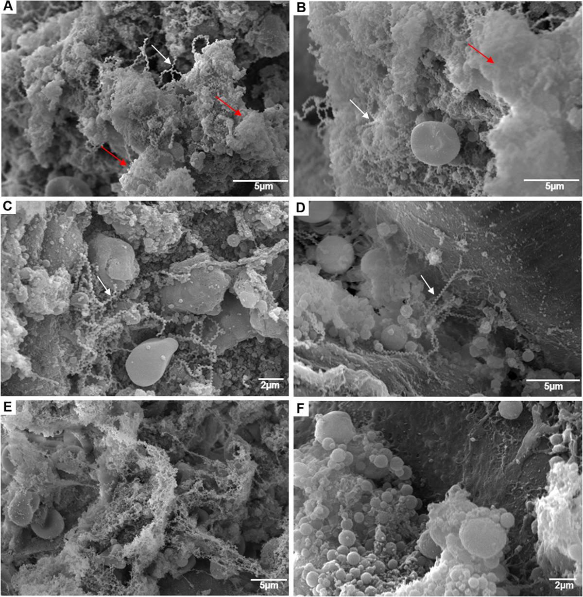
In reservoir hosts, leptospires colonize the kidney's proximal tubules and are shed in urine. Cellular details shown come from rodent studies .
Jim Miller has seen more feral hogs along the creek this year than ever.
For Pomona, cattle are incidental hosts: the source is usually another species .
Your turn
Check your understanding
1 question on this station · Score 0 / 1
Featured question
A 4-year-old Angus-cross cow aborts at 8 months of gestation after drinking from a creek. Feral swine use the upstream banks. Urine cultures from individually identified swine yield isolates typed as Pomona on three monthly visits. Those animals continue eating and moving with their groups.
Rat kidney, scanning electron microscopy: leptospire agglomerates in biofilm matrix inside tubules (natural rodent reservoir).
Santos AAN, et al. Leptospira interrogans biofilm formation in Rattus norvegicus natural reservoirs. PLoS Negl Trop Dis. 2021;15(9):e0009736, Fig. 2. · CC BY 4.0
Survival depends on conditions: leptospires persist in warm, moist soil and stagnant water (about 6 months, per one extension source) and die quickly when dried . Cold tolerance varies: some survived prolonged cold-water storage experimentally .
Heavy rain and flooding spread contaminated urine across pasture and water .
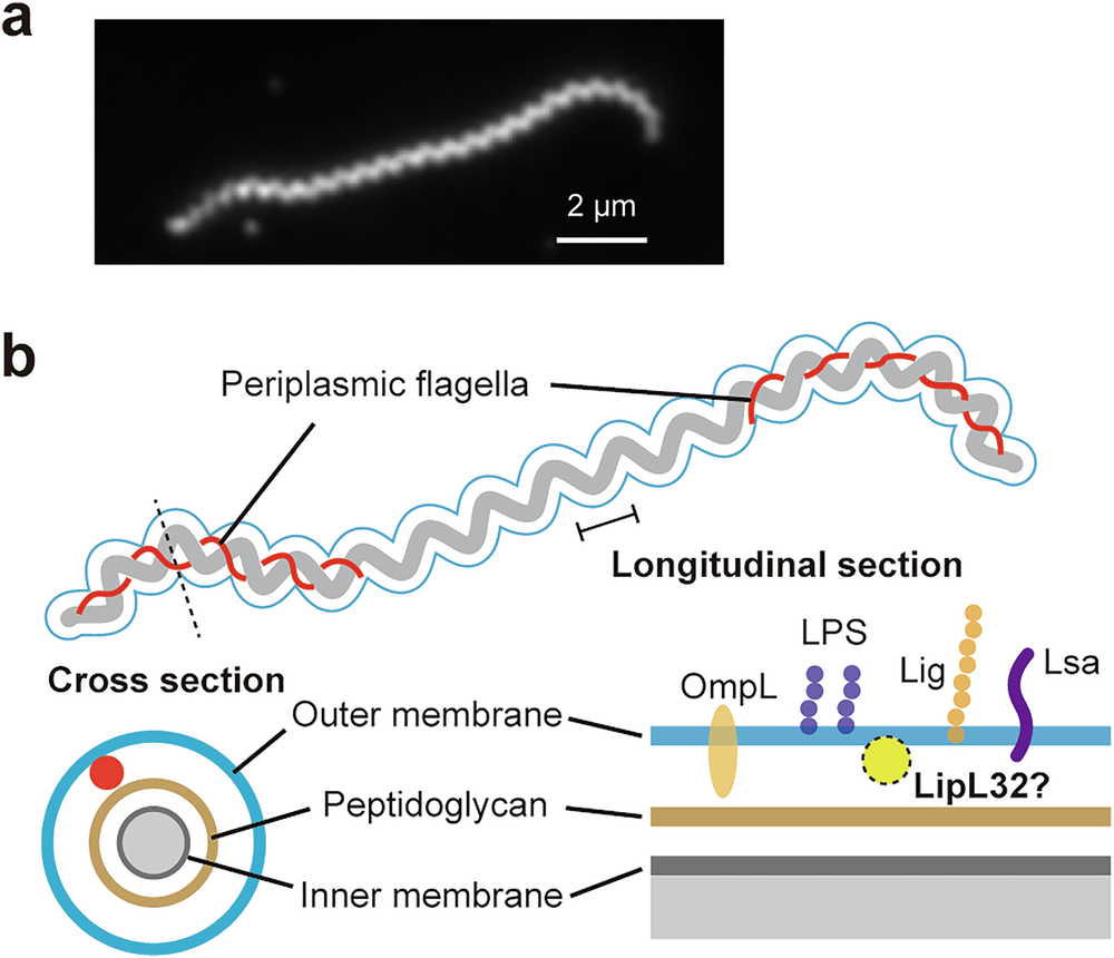
The periplasmic flagella lie between the outer and inner membranes near each end; they curve the cell ends and drive corkscrew motility .
A thin, helical spirochete with hooked ends, moving by internal (periplasmic) flagella; motility is essential for infection .
After weeks of rain, the pond edges are trampled mud.
Fence off stagnant water and muddy margins: removing the exposure is part of control .
Your turn
Check your understanding
1 question on this station · Score 0 / 1
Featured question
A 4-year-old Angus-cross cow belongs to a pasture herd with six abortions during the past five weeks. Feral swine were excluded ten days ago, and covered troughs now receive water from a separate well. During an afternoon inspection, this cow and eight herdmates have their fetlocks submerged beside the pond previously used by wildlife. Rodent droppings are present in the feed shed. Replacement heifers are due in three weeks, and the veterinarian has scheduled a herd vaccination review.
(a) Real dark-field image of Leptospira interrogans. (b) The authors’ diagram: periplasmic flagella lie between the outer and inner membranes near each end and curve the cell ends; LipL32’s surface exposure is uncertain (“LipL32?”).
Abe K, Koizumi N, Takazaki H, Hirose M, Takabe K, Kato T, Nakamura S. A periplasmic layer formed by an outer membrane lipoprotein governs the cell-envelope integrity and stiffness of Leptospira interrogans. Communications Biology, 2026, Fig. 1a. · CC BY 4.0
Schematic vs real
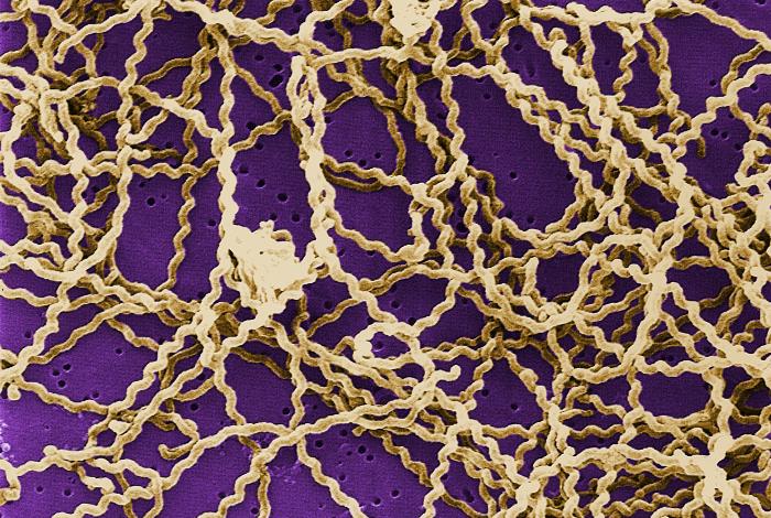
Scanning electron micrograph: the spiral shape of Leptospira.
CDC / Rob Weyant, Public Health Image Library #138. · Public domain
Schematic vs real
03 / 15Muzzle, eye, skin
Through the door
No wound needed: leptospires enter through mucous membranes or water-softened skin.
Day 0
Entry is through intact mucous membranes of the mouth or conjunctiva, or damaged or water-softened skin .
Motility helps leptospires penetrate tissue and spread .
Infection occurs by exposure of the mucous membranes of the eye, mouth, and genital tract .
Illustrative: the organism passes through tissue and reaches small vessels; the exact route in cattle is not established .
Cows drink and stand in the same muddy water.
People are infected the same way: the son's bare hands met fetal fluids .
Your turn
Check your understanding
1 question on this station · Score 0 / 1
Featured question
A 4-year-old Angus-cross cow has MAT results against Pomona of 1:100 and 1:800 fourteen days apart, without intervening vaccination. During the preceding week, she stood for six hours in floodwater reaching halfway up her metacarpal regions in a pen receiving runoff from affected cattle. Examination records describe a 1-cm abrasion on her flank and no visible breaks in the skin of her distal limbs. During the same week, she drank from a covered well-fed trough, rubbed her face against stored hay, and underwent artificial insemination. The veterinarian is reconstructing how the organism could have crossed an external barrier during the pen exposure.
04 / 15Lamina propria
The first alarm
Leptospires are 'stealthy': innate sensors see them poorly, so the early infection is often silent.
Day 0-2 (approximate)
Leptospiral LPS is atypical and less endotoxic than that of other Gram-negative bacteria; purified LPS signalled through TLR2 in human cells but TLR4 in mouse macrophages . Inactivated leptospires activated dog, mouse, and human TLR2 and mouse and human TLR4; TLR2 activity of purified LPS preparations was attributed to co-purified lipoproteins . Neither study used bovine cells; evidence from other species does not establish every step in cattle.
Live leptospires barely trigger TLR5; heat-killed bacteria activate bovine and human TLR5, because the flagellin (FlaB) is shielded inside the periplasm .
In bulls with natural genital infection, infection was associated with higher TLR2 expression in smegma (about 4-fold); TLR4 expression did not differ .
In animals, the acute phase is mostly subclinical; the main losses come from chronic infection and reproductive wastage .
A macrophage's TLR2 engages leptospiral lipoproteins; TLR4 recognition of LPS varies by host, and TLR5 stays largely silent while flagellin is hidden (schematic) .
The cows that aborted were never seen sick; only two heifers were briefly off feed.
Silent acute infection is why herd problems surface as abortions weeks later: plan herd-level testing, not just sick-animal workups .
Your turn
Check your understanding
1 question on this station · Score 0 / 1
Featured question
A 4-year-old Angus-cross cow is pregnant after grazing flooded pasture. Her Pomona MAT titre is 1:1600 at the first sampling and 1:6400 fourteen days later. No vaccination occurs between collections. She continues eating and walking with the herd throughout that interval.
05 / 15Circulation
The bloodstream
For roughly a week, leptospires multiply in blood and reach many tissues.
First days (approximate)
Leptospiremia lasts up to about 7 days and seeds many tissues .
Pathogenic leptospires recruit host complement regulators such as factor H, which reduces (but does not abolish) complement attack; this was shown mainly with human and horse serum systems, including serovar Pomona .
Fever and brief inappetence may be the only signs in adult cattle .
Complement proteins (purple) approach; factor H on the surface lowers their deposition, so most attacks fail.
Two heifers went off feed for a day or two.
This short bloodstream window is when the fetus is exposed .
Choose a path
Branch point: what kind of host is this cow for this serovar?
The tour waits for your choice.
Acute course: high, rising titers; abortion, stillbirth, weak calves; hemolysis in calves . This is our herd.
Mild or silent course: low titers, long renal and genital carriage, infertility and sporadic abortion .
Your turn
Check your understanding
1 question on this station · Score 0 / 1
Featured question
Five 2-year-old Angus-cross heifers from creek-water paddocks are evaluated during a herd abortion investigation. None has received antimicrobials, and one whole-blood PCR submission is available today for the bacterial target being investigated. Heifer 3 stopped eating nine days ago and has a rectal temperature of 40.2 °C today. Heifer 17 stopped eating fourteen days ago and has a rectal temperature of 40.4 °C today. Heifer 26 stopped eating twenty-one days ago and expelled an eight-month fetus this morning. Heifer 35 first entered a creek-water paddock yesterday, continues eating, and has a rectal temperature of 38.8 °C. Heifer 48 stopped eating yesterday and has a rectal temperature of 40.6 °C today.
06 / 15Endothelium
The vessel wall
Damage to small-vessel endothelium explains hemorrhages throughout the body.
First week (approximate)
Endothelial injury is a central lesion in severe leptospirosis; how much it explains each hemorrhage is not fully established .
Leptospires bind host matrix proteins (laminin, collagen, fibronectin) through several outer-membrane proteins .
Endothelial junctions loosen; red cells and plasma leak out (petechiae).
Hemorrhages may occur in fetuses aborted from incidental serovars .
Several proteins once called virulence factors (for example LipL32) proved dispensable in knockout studies: pathogenesis is multifactorial .
Your turn
Check your understanding
1 question on this station · Score 0 / 1
Featured question
A male Angus-cross bovine fetus is expelled at eight months of gestation after its dam grazed flooded pasture. Numerous red pinpoint spots cover the pleura and renal capsule. Histological sections show intact erythrocytes between collagen bundles surrounding capillaries at these sites. Clear fluid separates bundles of subcutaneous connective tissue and occupies the pleural cavity.
07 / 15Blood
Red cells and the calf
In calves with incidental serovars, red cells break open in the bloodstream.
First week (approximate) · in a calf
Leptospires produce hemolysins, including a sphingomyelinase (Sph2) and a separate pore-forming protein (SphH); their role in hemolysis in calves is not proven .
Calves with incidental serovars, especially Pomona, can develop severe disease: high fever, weakness, red urine, and rapid breathing from anemia .
Icterus and haemoglobinuria are especially seen in young animals .
Schematic: red-cell membranes are damaged and hemoglobin is released, then filtered into urine (hemoglobinuria).
One weak calf had dark red urine and breathed hard before it died.
Red urine could reflect hemoglobinuria from hemolysis. Confirm the mechanism with urinalysis and blood findings.
Your turn
Check your understanding
1 question on this station · Score 0 / 1
Featured question
A 10-day-old male Angus-cross calf was born fourteen days before its expected calving date during a herd abortion investigation. Its milk intake has declined over the past day. A freely voided urine specimen is centrifuged and examined within ten minutes; the supernatant is red, the pH is 6.5, and the specific gravity is 1.025. The sediment contains zero erythrocytes in ten examined high-power fields. Plasma separated promptly from two atraumatic jugular blood collections is pink.
08 / 15Hepatic cords
The liver
In a hamster model, leptospires slipped between liver cells and broke the bile canaliculi, letting bile leak into blood.
Approximate
In a hamster model, leptospires invaded the junctions between hepatocytes and destroyed bile canaliculi, causing jaundice .
Fetuses infected with Pomona may be icteric, but fetal lesions are not specific .
Tight junctions sealing the bile canaliculus open; green bile seeps into the sinusoid.
The aborted fetus showed mild icterus.
Mechanism shown is from a hamster model: an animal-model finding, not proven in cattle .
Your turn
Check your understanding
1 question on this station · Score 0 / 1
Featured question
A male Angus-cross bovine fetus at 8 months of gestation is submitted after the sixth abortion in five weeks. Yellow connective tissues are recorded during inspection. No organism assay has yet been performed on the submission. The dam's available serum was collected only at expulsion, and her last reproductive vaccine product is unknown.
09 / 15Bloodstream
The antibodies arrive
Agglutinating antibodies clear most leptospires from the blood; some persist in the kidney despite them.
From about day 6
Agglutinating and opsonizing antibodies appear around day 6 and clear leptospires from most organs .
Leptospires can persist in relatively protected sites such as the proximal renal tubules .
Immunity is largely antibody to leptospiral LPS and is serovar-specific; cross-protection is very little or absent .
A fourfold rise in titre between paired acute and convalescent sera indicates acute infection .
Y-shaped antibodies (blue) bind and clump leptospires (agglutination); phagocytes engulf the clumps.
In the laboratory, the MAT measured Pomona 1:1600, then 1:6400 two weeks later: a fourfold rise that supports recent infection; the serovar remains presumptive.
The MAT measures agglutination in serial dilutions of serum in the laboratory, not in the bloodstream.
Your turn
Check your understanding
1 question on this station · Score 0 / 1
Featured question
A 4-year-old Angus-cross cow aborts at 8 months of gestation. She received no reproductive vaccine during the preceding two years or between the current collections. Pomona MAT titres are 1:1600 at expulsion and 1:6400 fourteen days later, while Hardjo titres are 1:100 on both occasions. No fetal organism testing or isolate characterization has been performed.
10 / 15Diagnostic laboratory
What each test sees, and when
Each test captures one stage of infection; timing decides what you can find.
Day 0 to months
Blood (PCR or culture): useful only during the transient leptospiremia, up to about a week .
Serology (MAT): antibodies from about day 6; a fourfold rise in paired sera indicates acute infection; MAT cross-reacts between serovars; vaccinated animals may have titres to vaccine serovars .
Maintenance-host infections can show low titres: about one-third of cows aborting from Hardjo have titres below 1:100 .
Urine (PCR or culture): detects renal carriers; shedding is intermittent, so one negative sample does not exclude carriage; repeated weekly samples can strengthen evidence of non-shedding; follow the veterinarian's and laboratory's testing plan .
Fetus and placenta: fluorescent antibody, PCR, or immunohistochemistry on tissues; fetal antibodies are diagnostic but titres are often very low (around 1:10) .
Finding leptospires in the kidney, urine, or genital tract of a healthy animal shows carriage, not disease .
Timeline graphic: blood PCR window (first week) → MAT rising (from about day 6) → urine PCR (weeks to months, intermittent) → fetal tissues (at abortion).
This herd's workup used each window: fetal kidney PCR and FA, paired MAT, and urine PCR within 2 weeks of abortion.
Before interpreting any titre, ask: vaccinated? when? which vaccine?
Your turn
Check your understanding
1 question on this station · Score 0 / 1
Featured question
Five 4-year-old Angus-cross cows undergo follow-up during a herd abortion investigation, with today designated day 14. The same laboratory performs all urine PCR tests, and assay controls amplify as expected unless otherwise stated. Cow 18 has remained in a pen supplied with trough water since day −21, received a reproductive vaccine on day −7, has negative urine results on days 0, 7, and 14, and has Pomona MAT titres of 1:400 on day 0 and 1:1600 on day 14. Cow 52 has negative urine results on days −14, −7, and 0, returned to flooded pasture on day 1, and has no subsequent urine results. Cow 9 has negative urine results on days 0 and 7, but neither the bacterial target nor the internal amplification control produces a signal in today's specimen. Cow 36 has three negative urine specimens collected from separate voids today and no earlier results. Cow 63 has negative, positive, and negative urine results on days 0, 7, and 14, respectively.
11 / 15Pregnant uterus
The placenta and the fetus
In the brief bloodstream phase, leptospires can cross the placenta and kill the fetus.
During the bloodstream phase; abortion later (approximate)
Transplacental infection during the limited time of maternal leptospiremia is proposed as the cause of abortion; reproductive pathogenesis is still poorly understood .
Lesions: diffuse placentitis with avascular, light tan cotyledons and edematous, yellowish intercotyledonary areas; the fetus usually dies 1-2 days before expulsion and is autolyzed .
Non-Hardjo serovars (mainly Pomona, Grippotyphosa) cause late-term abortion, stillbirth, or premature weak calves .
A placentome is a maternal caruncle joined to a fetal cotyledon. Leptospires reach the placenta and fetus; placentitis follows. Exactly how the fetus dies is not fully understood .
Six late-gestation abortions in five weeks; fetal kidney PCR and FA positive.
By the time the fetus is expelled, the dam's titer may already be waning: test the fetus and placenta .
Your turn
Check your understanding
1 question on this station · Score 0 / 1
Featured question
A 4-year-old Angus-cross cow aborted at eight months of gestation three days ago. Her Pomona MAT titre was 1:6400 two weeks before expulsion and is 1:1600 today, with no vaccination between collections. The fetus remained outdoors without refrigeration until this visit, and its recovered kidneys separate into fluid and tissue fragments during handling. A veterinarian placed portions of the expelled placenta in neutral buffered formalin immediately after collection on the day of abortion. A screening section from that material shows cotyledonary epithelium with visible nuclei and aggregates of neutrophils. Maternal blood, serum, and urine can be collected today, and the receiving laboratory offers bacterial culture, PCR, MAT, and immunohistochemistry.
12 / 15Uterus, oviduct, and semen
The genital tract and the bull
In maintenance hosts, Hardjo colonises the genital tract: a silent cause of infertility, spread also by breeding.
Months to years (maintenance hosts)
Chronically infected animals can carry leptospires in the kidney and in the male and female genital tract .
Sejroe-serogroup leptospires (which include Hardjo) preferentially colonise the reproductive tract: ovaries, follicles, oviducts, uterus, cervicovaginal mucus, and semen .
Genital colonisation causes a chronic, silent, subclinical reproductive syndrome; Hardjo-infected cows are about twice as likely to fail to conceive .
Transmission may occur during mating; in one study 1.5% (4/268) of bull semen samples were PCR-positive and one yielded viable, infectious leptospires .
Recommended diagnosis: herd MAT screening, then PCR on genital samples .
Leptospires in cervicovaginal mucus and on the endometrial surface; in the bull, in semen (schematic).
This herd's problem is Pomona abortions, but its stable Hardjo titres (1:100) and three bulls are reasons to think about genital carriage too.
Repeat breeders and long calving intervals with low Hardjo titres? Consider genital leptospirosis and test genital samples by PCR .
Your turn
Check your understanding
1 question on this station · Score 0 / 1
Featured question
A 4-year-old Angus-cross cow stands to be mounted 21 days after each of two services by the same 4-year-old Angus-cross bull. Both animals continue eating their allotted feed throughout the breeding period. Neither has received a reproductive vaccine during the preceding year. Hardjo MAT titres are 1:100 in both animals on samples collected fourteen days apart. A urine specimen from each animal is lipL32 PCR-negative.
13 / 15Proximal tubule
The kidney
Hidden on the tubule brush border, leptospires are shed in urine and the cycle restarts.
Weeks in this herd; months to years in maintenance hosts
In mice, virulent leptospires reached the kidney, attached first to the basal side of tubular cells, then colonized the lumen and brush border .
In rat reservoirs they form aggregates and biofilm in proximal tubules ; in hamsters, some colonized tubules lacked inflammation, consistent with immune evasion .
Urine carries leptospires out. Urine PCR detects leptospiral DNA (not proof of live organisms); for non-Hardjo serovars, test urine within 2 weeks after abortion .
Aggregates on the brush border; microvilli disorganize; leptospires detach into tubular fluid and leave in urine.
Urine PCR positive in 2 of 5 aborting cows: these cows may be shedding into the pond.
Shedding is intermittent: one negative urine does not clear a cow .
Your turn
Check your understanding
1 question on this station · Score 0 / 1
Featured question
A 4-year-old Angus-cross cow enters follow-up after grazing flooded pasture. On day zero, blood PCR targeting pathogenic Leptospira is positive and her Pomona MAT titre is 1:100. From that day onward, she occupies an individual pen with piped drinking water, protected feed, and no breeding or contact with other animals. Blood PCR is negative on days 14 and 28, and her day-14 Pomona MAT titre is 1:6400. Urine collected by sterile urethral catheterization after vulvar cleansing on days 28 and 42 yields isolates identified as the same serovar. She receives neither vaccination nor antimicrobials during this interval.
14 / 15Herd
Breaking the cycle
Control combines treatment, serovar-matched vaccination, and cutting exposure to water and wildlife.
Now and every year
Immunity is largely serovar-specific; cross-protection is very little or absent, so multivalent vaccines are used .
Most cattle reproductive vaccines include an L5 component (Canicola, Grippotyphosa, Hardjo, Icterohaemorrhagiae, Pomona); some add Hardjo-bovis .
Hardjo vaccines reduce urinary shedding (pooled efficacy 88.7% in a meta-analysis); in one challenge trial a monovalent Hardjo-bovis vaccine prevented renal colonisation and shedding, but protection is not sterile across all products .
Heifers: two doses 4-6 weeks apart before breeding, then annual boosters . Treat infected cattle before vaccination is expected to work .
Treatment options include oxytetracycline, tilmicosin, ceftiofur, and amoxicillin; some uses are extralabel and must follow FDA rules (cephalosporins only at approved dose, frequency, duration, and route) .
Environment: fence stagnant water and muddy margins, prevent urine contamination of feed, control rodents and feral swine .
Herd diagram: vaccine-induced antibodies (blue) meeting leptospires at entry; fewer reach the kidney; fewer in urine.
Jim Miller's plan: treat under veterinary direction, vaccinate the whole herd, fence the pond margins, provide clean water, and work on feral hog control.
Vaccination changes serology: record products and dates so future titres can be interpreted .
Your turn
Check your understanding
1 question on this station · Score 0 / 1
Featured question
A 4-year-old Angus-cross cow is followed during a herd abortion investigation. Her records document completion of the labelled primary vaccination course and a scheduled booster four weeks before follow-up sampling. Since the investigation began, she has occupied a paddock supplied with piped drinking water and separated from the pond by fencing. Her follow-up urine culture yields an isolate that the reference laboratory identifies as a serovar included in her vaccine.
15 / 15The farm family
People at the pond
Leptospirosis is a zoonosis: farm families, veterinarians, and workers share the cattle's exposure.
Any time
People at risk include veterinarians and veterinary staff, people raising farm animals, dairy workers, and slaughterhouse workers .
Prevention: avoid exposure; when unavoidable, wear rubber boots, waterproof clothing, gloves, and eye protection, cover cuts, and wash hands .
An estimated 1 million people are affected worldwide each year .
A person with symptoms after exposure needs prompt medical evaluation and should mention the animal exposure; diagnosis and treatment belong to the physician .
Hands, eyes, and small cuts as entry points; PPE icons closing each one.
The son had fever and aches after handling fetuses bare-handed: he should see his physician promptly and mention the cattle.
Veterinarians are often the first to recognise a zoonotic risk on a farm: say it clearly and recommend medical care.
Your turn
Check your understanding
1 question on this station · Score 0 / 1
Featured question
On October 12, a 4-year-old Angus-cross cow aborts at eight months of gestation. The owner's adult son assists without gloves, and fetal fluids cover fissures across his knuckles. Fetal kidney lipL32 PCR is positive. On October 19, he develops headache and calf muscle pain and records an oral temperature of 39.2 °C; a medical appointment is arranged for October 22. His history also includes freshwater swimming on August 20. A household member developed a cough on October 17. He saw a tick attach to his forearm during an October 20 hike and removed it that day. On October 21, cattle urine splashed into his eye during pen cleaning.